Introduction to Cardiac Contractility
Cardiac contractility refers to the intrinsic ability of the myocardium to contract, independent of preload (ventricular filling) and afterload (arterial pressure). Assessing contractility is vital in both clinical cardiology and cardiovascular research, as it provides insight into myocardial health, response to drugs, and the progression of diseases like heart failure.
Two of the most recognized measures for quantifying ventricular contractility are dP/dt max and the End-Systolic Pressure-Volume Relationship (ESPVR). Although both are linked to contractile performance, they differ in methodology, interpretation, and sensitivity to external influences.
Overview of dP/dt max
Definition and Physiological Basis of dP/dt max
The term dP/dt max represents the maximum rate of pressure rise during ventricular contraction — essentially, how quickly the left ventricular pressure increases during systole. It’s derived from the ventricular pressure waveform, typically measured via high-fidelity micromanometers.
Mathematically, it’s expressed as:
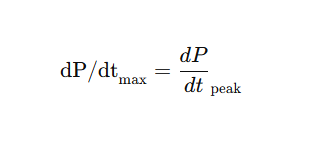
where P is pressure and t is time.
This measurement reflects the contractile velocity of myocardial fibers and is often used as a rapid, sensitive index of contractility in both clinical and laboratory settings.
How dP/dt max Is Measured
dP/dt max is obtained from left ventricular pressure tracings using invasive catheters. It can also be estimated noninvasively using echocardiography, though such methods are less precise.
Advantages and Limitations of dP/dt max
Advantages:
- Easy and fast to measure.
- Useful in real-time hemodynamic monitoring.
- Sensitive to inotropic interventions (e.g., dobutamine, digoxin).
Limitations:
- Highly dependent on preload and afterload.
- Doesn’t fully isolate intrinsic myocardial contractility.
- Can vary significantly with heart rate and arrhythmias.
Understanding ESPVR (End-Systolic Pressure-Volume Relationship)
Definition and Concept Behind ESPVR
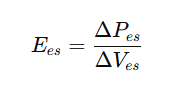
ESPVR represents the relationship between end-systolic pressure and volume during different loading conditions. When plotted on a pressure-volume loop, the end-systolic points form a straight line — the end-systolic pressure-volume line (ESPVL).
The slope of this line (Ees) is a load-independent index of contractility, where:
The Role of ESPVR in the Pressure-Volume Loop
In the pressure-volume (PV) loop, ESPVR defines the maximum pressure the ventricle can generate at a given volume. A steeper slope indicates stronger contractility, while a flatter slope suggests impaired contractility.
How ESPVR Reflects Ventricular Contractility
ESPVR is considered more physiologically accurate because it’s less sensitive to preload and afterload, offering a clearer view of the heart’s intrinsic pumping ability.
dP/dt max versus ESPVR: Key Differences and Comparative Analysis
| Feature | dP/dt max | ESPVR |
| Definition | Max rate of pressure rise during systole | Slope of end-systolic pressure-volume line |
| Sensitivity to Load | Load-dependent | Load-independent |
| Ease of Measurement | Simple and quick | Requires full PV loop analysis |
| Physiological Meaning | Reflects contractile speed | Reflects intrinsic contractility |
| Best Use Case | Acute monitoring | Comprehensive cardiac assessment |
| Accuracy | Moderate | High |
| Common Application | ICU and cath lab | Research and advanced diagnostics |
While dP/dt max is ideal for real-time functional assessment, ESPVR remains the gold standard for quantifying intrinsic contractility.
Clinical Applications and Diagnostic Relevance
Use of dP/dt max in Clinical and Experimental Settings
In critical care, dP/dt max provides a quick and dynamic reflection of ventricular performance. It’s valuable during:
- Inotropic drug titration
- Mechanical support adjustments (e.g., LVADs)
- Hemodynamic monitoring during surgery
Use of ESPVR in Advanced Cardiac Studies
ESPVR, by contrast, is primarily used in experimental physiology and advanced imaging studies, such as conductance catheterization. It helps evaluate the true contractile function independent of external loading influences.
Experimental and Computational Insights
Role of Pressure-Volume Loop Analysis in Research
The pressure-volume (PV) loop is a powerful graphical representation of the cardiac cycle. It helps visualize changes in pressure (P) and volume (V) during each heartbeat. By altering preload and afterload conditions experimentally, researchers can derive both dP/dt max and ESPVR to evaluate the ventricular contractility curve.
PV loops provide a comprehensive picture of ventricular performance, integrating parameters like stroke volume, cardiac output, end-systolic pressure, and relaxation rate.
- dP/dt max reflects the steepness of the upstroke, highlighting the rapid pressure build-up phase.
- ESPVR, on the other hand, characterizes the upper-left boundary of the loop, indicating the maximum pressure the ventricle can generate for a given end-systolic volume.
Together, they offer complementary insights — dP/dt max for instantaneous contractile function, and ESPVR for steady-state contractility.
Integration of Both Metrics in Cardiac Modeling
Modern computational models of cardiac mechanics often combine both dP/dt max and ESPVR to simulate ventricular dynamics under varying physiological or pathological states.
This integration allows for:
- Predictive modeling of heart failure progression.
- Simulation of inotropic interventions and their hemodynamic effects.
- Personalized cardiac diagnostics, aiding in precision medicine.
Advanced simulation tools and finite element modeling can even adjust for load-dependence and nonlinearities, refining both indices for clinical translation.
Limitations and Technical Challenges
Challenges in Measuring dP/dt max Accurately
Despite its clinical convenience, dP/dt max comes with measurement pitfalls:
- It’s highly preload-dependent — meaning increased filling pressure can falsely elevate values.
- Afterload changes (like hypertension) can also distort results.
- Catheter position and signal filtering influence the accuracy of waveform-derived measurements.
- In noninvasive echocardiographic methods, image quality and Doppler alignment can limit reliability.
Therefore, clinicians often interpret dP/dt max in context, considering other hemodynamic variables like LV end-diastolic pressure (LVEDP) or ejection fraction.
Limitations in ESPVR Estimation and Linear Assumptions
While ESPVR is considered load-independent, it’s not entirely exempt from variability:
- The linear assumption of the ESPVR line may not hold under extreme physiological conditions.
- Obtaining accurate end-systolic points requires multiple cardiac cycles under variable loading, making it technically demanding.
Summary Table: dP/dt max vs ESPVR Comparison
| Parameter | dP/dt max | ESPVR |
| Definition | Max slope of LV pressure rise during systole | Slope of end-systolic pressure-volume relationship |
| Load Dependence | Load-dependent | Largely load-independent |
| Measurement Method | Pressure catheter / echocardiography | Conductance catheter / PV loop |
| Data Required | Single pressure waveform | Multiple PV loops under varied loading |
| Clinical Utility | Quick functional assessment | Comprehensive contractility analysis |
| Accuracy | Moderate | High (gold standard) |
| Ease of Use | Simple and fast | Complex and invasive |
| Application | ICU, pharmacological testing | Research, advanced cardiology |
| Interpretation | Reflects contractile speed | Reflects intrinsic contractile strength |
Frequently Asked Questions (FAQs)
1. What does dP/dt max actually measure in the heart?
dP/dt max measures the maximum rate of left ventricular pressure rise during early systole. It indicates how quickly the ventricle can build pressure, reflecting myocardial fiber shortening velocity.
2. Why is ESPVR considered more accurate than dP/dt max?
ESPVR is less influenced by preload and afterload, making it a more reliable indicator of the heart’s intrinsic contractility. It isolates contractile function without being heavily affected by external conditions.
3. Can dP/dt max and ESPVR be used together?
Yes. Using both provides a comprehensive assessment — dP/dt max captures real-time functional changes, while ESPVR gives a load-independent baseline of myocardial performance.
4. Is dP/dt max reliable for patients with heart failure?
In heart failure, dP/dt max may underestimate contractility due to altered loading conditions and compliance changes. Combining it with ESPVR or ejection fraction yields more accurate insights.
5. How is ESPVR measured in humans?
ESPVR is typically obtained using conductance catheters, requiring controlled loading variations.
6. Which is better for drug testing — dP/dt max or ESPVR?
For acute drug response studies (e.g., evaluating inotropes), dP/dt max is preferred for its rapid responsiveness. For mechanistic or dose-response modeling, ESPVR offers deeper physiological understanding.
Conclusion: Integrating Both Metrics for Comprehensive Cardiac Assessment
In the debate of dP/dt max versus ESPVR, the answer isn’t about superiority but complementarity.
- dP/dt max excels in speed, simplicity, and real-time feedback, making it indispensable in clinical and critical care settings.
- ESPVR, with its load-independent precision, remains the gold standard for understanding the heart’s intrinsic contractile mechanics.
For researchers, integrating both metrics provides a multi-dimensional understanding of ventricular function, blending temporal precision with mechanical accuracy.
Ultimately, both serve as vital instruments in the broader symphony of cardiac performance assessment — each playing its role in advancing cardiovascular science and patient care.