Structural Heart Interventions
With the increasing development of various structural heart devices, CD Leycom aims to incorporate real-time PV loop hemodynamics to enhance the intra-operative experience of structural heart interventions and improve patient outcomes.
Mitral Valve Clipping
Mitral regurgitation (MR) is the most common form of valvular heart disease. Mitral valve clipping is a catheter based procedure that provides a less invasive alternative for surgical mitral valve repair. In this procedure, one or more metal clips are deployed to create a double orifice mitral valve by clipping the leaflets together to reduce MR.
It may therefore shorten the procedure by the real-time evaluation of acute decreases in mitral regurgitation, eventually avoiding the placement of more than one clip.
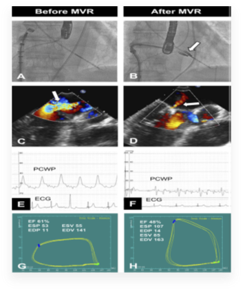
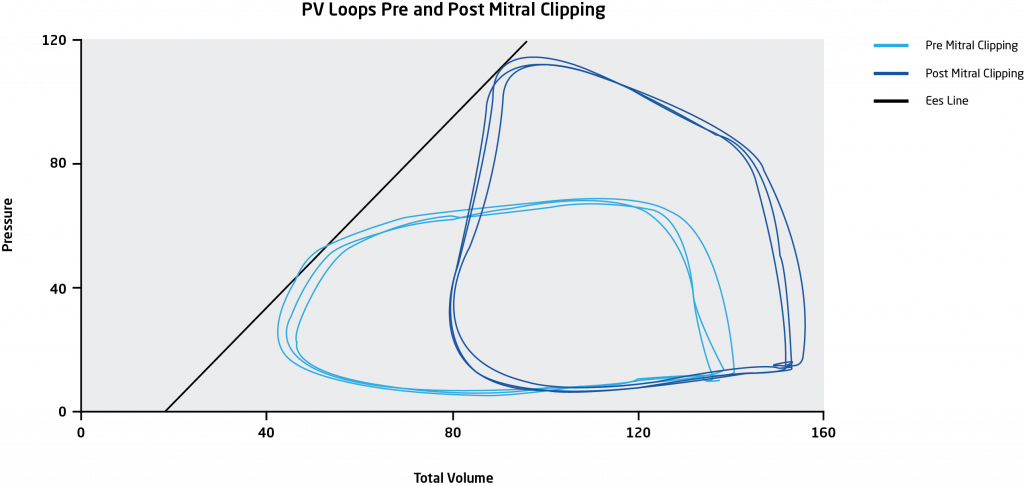
Clinical data by Gaemperli et al.1 demonstrated the use of pressure-volume (PV) loops to provide real time monitoring of the hemodynamic effects of MR reduction in the LV during mitral valve clipping. In their Clinical Perspective, the authors stated, “Real-time pressure-volume measurements may be used to monitor immediate changes in LV loading conditions and contractility during the MitraClip procedure, particularly in high-risk patients with low ejection fraction.”
The example below shows the acute improvement in LV function following the MitraClip procedure according to the end-systolic pressure-volume relationship.
Transcatheter Aortic Valve Replacement (TAVR/TAVI)
Transcatheter aortic valve replacement (TAVR), also known as transcatheter aortic valve implantation (TAVI), has become a common alternative to surgical aortic valve replacement for patients with severe aortic valve stenosis and a high level of surgical risk.
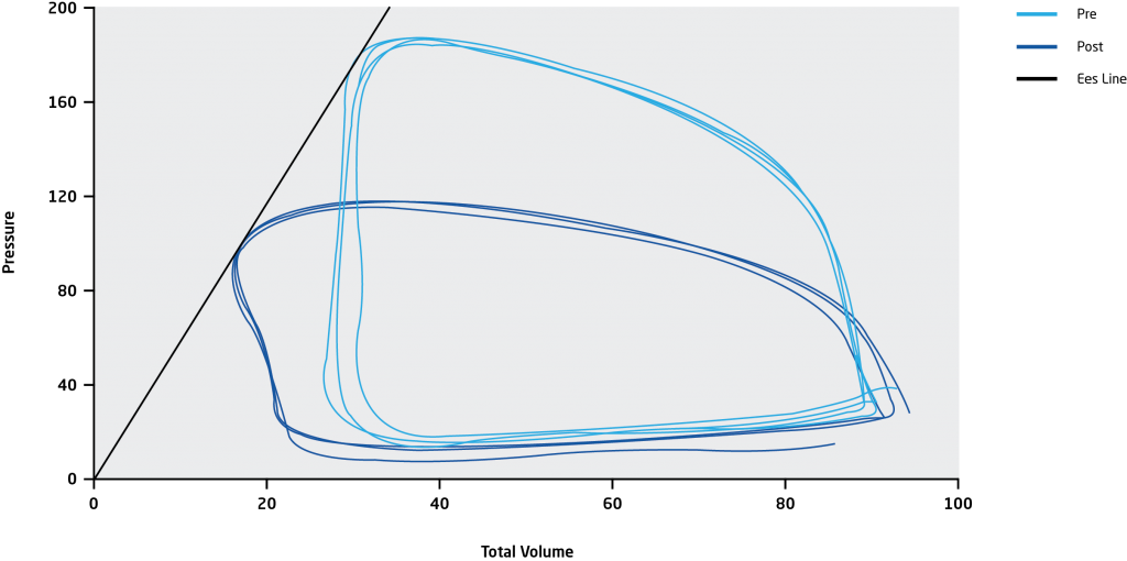
However, 20% of TAVR implants show paravalvular leakage, which is difficult to detect accurately with echocardiography.2 Using pressure-volume (PV) loop monitoring, this leakage can be detected and better quantified during the isovolumetric relaxation phase. Moreover, acute paravalvular leakage will immediately shift the left ventricular PV loop plane to the right due to acute volume overloading.
The newest generation of percutaneous valves can be repositioned mid-procedure, which in combination with PV analysis, may result in optimal valve placement with minimal or no paravalvular leakage.
Related publications.
- 2020; LV Hemodynamic Changes During TAVR Assessed By Real-Time PV Loops: https://interventions.onlinejacc.org/content/13/18/2190
- 2019; PV Loop Effects of TAVR for Moderate Aortic Stenosis with Reduced LV Ejection Fraction: https://interventions.onlinejacc.org/content/12/7/684
- 2019; PV Loop Analysis of Paradoxical Severe Aortic Stenosis: https://link.springer.com/article/10.1007/s00392-019-01423-z
- 2013; Real-Time LV PV Loops With MitraClip System: https://www.ahajournals.org/doi/10.1161/CIRCULATIONAHA.112.135061