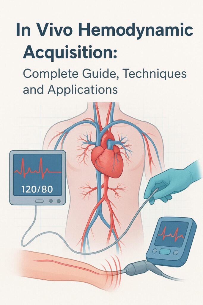
Introduction to In Vivo Hemodynamic Acquisition
Understanding how blood flows, how the heart pumps, and how vessels respond in real-time is critical for modern medicine. This process, known as in vivo hemodynamic acquisition, allows scientists and clinicians to measure, monitor, and analyze cardiovascular function directly within living organisms.
By gathering real-time data, researchers can investigate disease mechanisms, evaluate treatment outcomes, and optimize surgical procedures. With cardiovascular diseases ranking as the leading cause of mortality worldwide, the ability to acquire accurate hemodynamic measurements is more important than ever.

Defining Hemodynamics and Its Clinical Importance
Hemodynamics refers to the study of blood flow and circulation within the cardiovascular system. Key aspects include:
- Cardiac output – the volume of blood pumped by the heart per minute.
- Blood pressure – the force exerted by circulating blood against vessel walls.
- Vascular resistance – the opposition faced by blood as it flows through vessels.
Precise assessment of these parameters is vital in diagnosing and managing conditions such as hypertension, heart failure, coronary artery disease, and stroke.
What Does “In Vivo” Mean in Biomedical Research?
The term in vivo comes from Latin, meaning “within the living.” In biomedical contexts, it indicates that measurements or experiments are performed inside a living organism—unlike in vitro (in a controlled environment outside the body) or ex vivo (outside the organism but using living tissues).
This distinction matters because in vivo methods capture the true physiological state, accounting for biological variability, systemic regulation, and environmental influences that cannot be replicated in vitro.
Principles of Hemodynamic Measurements
Blood Flow Dynamics and Pressure Regulation
Blood flow is influenced by multiple physiological factors, including:
- Heart contractions generating pressure gradients.
- Elasticity of vessels, which allows smooth distribution of blood.
- Autonomic nervous system regulation, controlling vessel dilation and constriction.
Key Parameters in Hemodynamic Studies
Researchers typically measure:
- Systolic and diastolic pressure
- Pulse wave velocity (PWV)
- Stroke volume (SV)
- Peripheral resistance
- Oxygen saturation and perfusion rates
Collectively, these parameters give a detailed view of cardiovascular performance.
Techniques for In Vivo Hemodynamic Acquisition
Invasive vs. Non-Invasive Measurement Approaches
- Invasive methods: Use catheters or sensors inserted into vessels or heart chambers for highly precise measurements.
- Non-invasive methods: Employ imaging and external sensors, offering lower risk and better patient comfort.
Catheterization Techniques
Cardiac catheterization remains the gold standard for invasive hemodynamic measurement. It enables:
- Direct measurement of intracardiac pressures
- Blood sampling for oxygen content analysis
- Calculation of cardiac output using thermodilution
Doppler Ultrasound Applications
Doppler ultrasound is widely used for non-invasive blood flow assessment. It measures:
- Flow velocity in arteries and veins
- Valvular function in cardiac imaging
- Hemodynamic changes during stress tests
Magnetic Resonance Imaging (MRI) in Hemodynamics
MRI offers a non-invasive and highly accurate technique for visualizing hemodynamics. It is particularly useful for:
- Mapping blood flow patterns in complex vascular structures
- Detecting congenital heart defects
- Quantifying tissue perfusion
Optical Imaging and Photoacoustic Methods
Emerging imaging modalities such as optical coherence tomography (OCT) and photoacoustic imaging allow researchers to observe microvascular hemodynamics with remarkable resolution.
Data Acquisition Systems and Processing
Sensors and Transducers for Hemodynamic Monitoring
Modern hemodynamic acquisition relies on:
- Pressure transducers for direct vessel monitoring
- Flow probes for cardiac output measurement
- Wearable biosensors for continuous monitoring
Signal Processing and Noise Reduction
Raw hemodynamic data often contains noise due to movement artifacts and biological variability. Advanced algorithms are applied for:
- Filtering signals
- Improving accuracy
- Synchronizing multi-sensor data
Software Tools for Data Analysis
Software platforms such as LabChart and MATLAB-based toolkits help researchers visualize, process, and analyze hemodynamic datasets with greater precision.
Applications of In Vivo Hemodynamic Acquisition
Cardiovascular Disease Research
From hypertension to atherosclerosis, hemodynamic data plays a pivotal role in:
- Understanding pathophysiology
- Evaluating treatment outcomes
- Developing predictive models
Neuroscience and Cerebral Hemodynamics
Monitoring cerebral blood flow is essential for conditions like:
- Stroke
- Traumatic brain injury (TBI)
- Neurodegenerative disorders
Pharmacological Studies and Drug Development
In vivo hemodynamics allow scientists to test how new drugs affect cardiovascular performance, ensuring both safety and efficacy.
Surgical and Interventional Planning
Cardiac surgeons and interventional radiologists rely on hemodynamic data to:
- Plan stent placements
- Guide bypass procedures
- Evaluate post-operative outcomes
Challenges and Limitations
Biological Variability in Measurements
- Age, gender, and lifestyle factors introduce variability in results.
- Reproducibility can be challenging across populations.
Technical Constraints and Calibration Issues
- Catheter-based methods carry risks of infection.
- Sensor drift and calibration errors may compromise accuracy.
Ethical Considerations in Animal and Human Studies
- Animal models provide valuable insights but raise ethical debates.
- Human trials must balance risk vs. benefit carefully.
Future Trends in Hemodynamic Research
AI and Machine Learning in Hemodynamic Data Analysis
Machine learning algorithms are being trained to:
- Predict cardiovascular events
- Personalize treatment strategies
- Automate data interpretation
Miniaturized and Wearable Monitoring Devices
Wearables that continuously track blood pressure, heart rate, and vascular stiffness will revolutionize long-term monitoring.
Personalized Medicine and Predictive Analytics
By integrating genomics, lifestyle data, and hemodynamics, clinicians can tailor therapies to individual patients.
FAQs on In Vivo Hemodynamic Acquisition
Q1. What is in vivo hemodynamic acquisition used for?
It is used to measure cardiovascular function, blood flow, and pressure inside living organisms for research, diagnostics, and treatment planning.
Q2. Is in vivo measurement better than in vitro?
Yes, because it captures real physiological conditions that in vitro studies cannot fully replicate.
Q3. What are the most common non-invasive methods?
Doppler ultrasound and MRI are the most widely used non-invasive approaches.
Q4. Are invasive techniques safe?
They provide precise results but come with risks such as bleeding, infection, or vessel damage.
Q5. How does AI impact hemodynamic research?
AI enhances data analysis, reduces human error, and enables predictive modeling in cardiovascular studies.
Q6. Can wearable devices measure hemodynamics accurately?
Emerging wearables are becoming increasingly accurate, especially when combined with advanced signal processing.
Conclusion: The Future of Hemodynamic Insights
In vivo hemodynamic acquisition stands at the intersection of physiology, engineering, and clinical practice. It has already transformed our understanding of cardiovascular health and continues to evolve with new imaging technologies, AI-driven analytics, and wearable devices.
As methods become less invasive, more accurate, and widely accessible, the future holds the promise of personalized, real-time cardiovascular monitoring, improving outcomes for millions worldwide.
